Radiology Room
Panoramic X-ray, Cone beam scanner, X-ray of the TMJ and sinuses
Radiology Room
Panoramic X-ray, Cone beam scanner, X-ray of the TMJ and sinuses
Fully equipped!
Our Clinic is equipped with the latest cone beam dental scanner from the brand MyRay
The cone beam computed tomography (CBCT) technology in dentistry is a medical imaging technique that uses a cone-shaped X-ray beam to create detailed three-dimensional images of the maxillofacial region. These scanners are specifically designed to capture accurate images of the bony structure, teeth, soft tissues, and airways, making them extremely useful in many areas of dentistry, including implantology (dental implants), orthodontics (ODF), oral surgery, and endodontics.
Frequently Asked Questions (FAQ)
The indications for performing a dental cone beam are increasingly numerous and include :
- Preparation for dental implant placement
- Periodontal diseases
- Bone loss
- Assessment of an endodontic pathology
- Major oral surgery cases
- Investigation for a foreign body
- Evaluation of bone lesions
- Localization of nerves for wisdom tooth surgery
- Assessment of facial sinus pathologies
- Dental cysts
- Impacted teeth
- Pathologies of the temporomandibular joint (TMJ)
- Assessment of an infectious or tumoral pathology
Its advantages include high accuracy, three-dimensional visualization, reduced radiation exposure, rapid image acquisition, and precise diagnosis of dental and maxillofacial issues. In summary, CBCT is an essential tool for the precise planning of dental treatments and the reduction of risks for patients.
Exams available at the clinic:

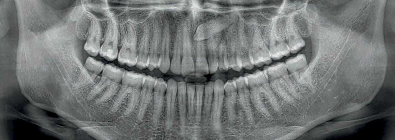
PANORAMIC X-RAY
The dental panoramic provides a complete view of the dentition and surrounding bony structures in a single radiographic image.

TMJ EXAMS
(OPEN OR CLOSED MOUTH)
The examinations of the temporomandibular joints (TMJ) assess the function, structure, and any potential pathologies of these crucial joints for chewing and speech.

EXAMINATION OF THE MAXILLARY SINUSES
The examinations of the maxillary sinuses allow for the assessment of the health and structure of these air cavities located in the facial region, often performed using X-rays or scans to diagnose conditions such as sinusitis or polyps.

ODONTOLOGICAL EXAMINATIONS

ORTHOPEDIC EXAMINATIONS
2D-3D Clinical Cases
Orthopantomograms (OPG)
- Digital "bitewing" radiographs: They allow for checking the condition of dental crowns with very good precision while using a low dose of radiation. It is an excellent alternative to traditional X-rays in the mouth, especially for patients with a strong gag reflex.
- Orthogonal panoramic: This type of image reduces overlaps between the teeth, making it easier to analyze the health of the gums and the bone that supports them.

Advanced Implant Planning
Thanks to modern tools, we can visualize your mouth in 3D and choose the ideal location for placing the dental implant. We also use very precise images taken with a scanner, which allows us to carefully plan your future smile. Everything is done to ensure a safe implant placement, respecting sensitive areas such as the jaw nerves.

Endodontic investigation
Mandibular canal therapy, identification of microfractures and root resorptions; the exceptional resolution of 68 μm, exclusive to Hyperion X9 pro, elevates your diagnostics to a higher level

Volumetric analyses
The function of the maxillary sinus lift volume calculation software allows for pre-determining the intervention and operating safely. Additionally, it is possible to draw lines directly on the patient's virtual model while assessing the morphological relationships on the 3D rendering.

The images used are the property of their respective owners MyRay ©
Simplified appointment booking!
Vous pouvez dès maintenant réserver votre rendez-vous en ligne !
Simplified appointment booking!
You can now book your appointment by clicking the button.